Homocysteine as Both a Vascular and Inflammatory Risk Marker
Elevated levels damage vessel walls and reprogram immune cells through two independent mechanisms.

Most cardiovascular biomarkers tell you where damage has already happened. LDL particles, troponin, even calcium scores: these are downstream readouts, numbers that move after a process is underway. Homocysteine works differently. It operates upstream, producing the damage that other markers later pick up. That's an unusual position for a blood test to occupy, and it's the reason homocysteine deserves its own article rather than a line in a cardiovascular risk panel.
Homocysteine is a sulfur-containing amino acid, made as a byproduct of methionine metabolism. Nobody eats it and nobody manufactures it on purpose. The body generates it constantly as a matter of ordinary chemistry, then recycles or clears it through pathways that depend on specific nutrients. When those pathways slow down, homocysteine accumulates in the blood, a state called hyperhomocysteinemia. Clinically it's graded as mild (15 to 30 µmol/L), moderate (30 to 100 µmol/L), or severe (above 100 µmol/L), and mild-to-moderate elevation appears in a meaningful share of the general population.
Homocysteine earns a closer look, but not just because elevated levels track with cardiovascular disease. Two separate, mechanistically documented pathways explain how it drives that disease: direct injury to the vessel wall, and a systemic inflammatory signal that operates through a different set of cells. Few biomarkers can claim both, and that dual action is what makes the rest of this piece worth working through.
How homocysteine physically damages the vessel wall
The vascular injury pathway is the better-studied of the two, and it doesn't rely on a single mechanism. Several things happen at once, converging on the same stretch of tissue: the endothelium, the thin layer of cells lining every blood vessel.
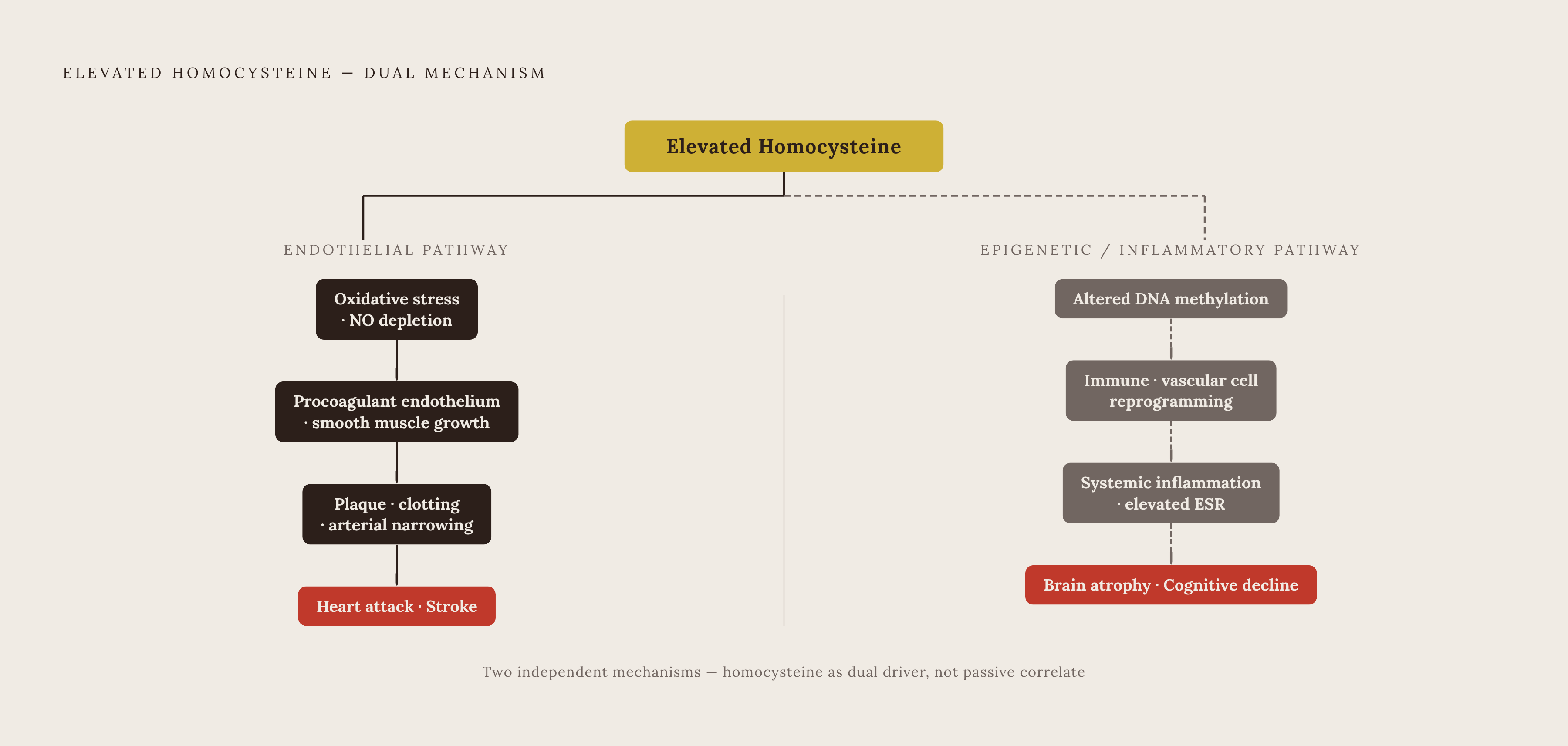
The clearest and most clinically relevant shift is functional. A 2025 review in the International Journal of Molecular Sciences describes how elevated homocysteine converts the endothelium from an anticoagulant surface into a procoagulant one. Healthy endothelium resists clot formation. Under sustained homocysteine exposure, it starts promoting it: coagulation factors get activated, and the normal balance between vasodilators and vasoconstrictors breaks down. The combined effect tilts the vessel wall toward clot formation and arterial narrowing, the two physical events behind most heart attacks and strokes.
Oxidative stress drives much of this. Homocysteine generates reactive oxygen species that deplete nitric oxide, the molecule responsible for relaxing blood vessels. Less nitric oxide means less vasodilatory capacity and a more permeable endothelial layer, conditions that invite further damage. Alongside the oxidative stress, homocysteine produces protein homocysteinylation, DNA damage, and apoptosis in endothelial cells, each a documented toxic effect in its own right.
The damage doesn't stop at the endothelial surface. Research published in PMC found that homocysteine directly activates the angiotensin II type 1 receptor, displacing angiotensin II itself. That matters beyond atherosclerosis: it implicates homocysteine in blood pressure dysregulation and aneurysm risk, mechanisms separate from plaque buildup. And in vascular smooth muscle cells, a study published in PMC found that homocysteine induces proliferative and migratory behavior, pushing these cells toward a foam-cell-like state. That's a direct contribution to plaque formation, not a passive byproduct of other damage. Four mechanisms, one target: the vessel wall takes a coordinated assault, which helps explain why even modest elevation can carry outsized risk.
How homocysteine drives systemic inflammation through a separate pathway
The inflammatory pathway runs on its own track. It runs independently of the endothelial mechanisms described above, and that independence makes homocysteine a genuine dual-pathway marker.
This arm runs mostly through immune cells and epigenetic change, a cast of characters you don't see in the coagulation factors and smooth muscle cells of the vascular story. Hyperhomocysteinemia drives changes in DNA methylation, and that shifts how genes express in immune and vascular cells. That's a slower, structural kind of damage. Elevated homocysteine doesn't just provoke a short-term inflammatory flare. It can reprogram how cells behave over time, altering their inflammatory posture well past the point where homocysteine exposure first occurred.
Clinical data backs this up outside the lab dish. A 2025 comparative study in ACS Omega found that elevated homocysteine, particularly in cardiac and diabetic patients, correlates with erythrocyte sedimentation rate, a standard marker of systemic inflammation. So the inflammatory arm operates in real patients, not only in cultured cells under controlled conditions. Put the two pathways side by side: one wears down the physical structure of the vessel wall, the other reprograms immune and vascular cells at a genetic level. They're distinct processes with distinct evidence bases, and that distinctness is the whole case for treating homocysteine as a dual-pathway marker.
What the two pathways mean for risk across organs
Vascular injury and inflammatory signaling don't confine themselves to coronary arteries. Both operate wherever blood vessels and immune cells exist. That means both operate in the brain just as readily as in the heart. That reach is what explains why homocysteine keeps turning up across disease categories that, on the surface, have nothing to do with each other.
The brain data makes this concrete. The Framingham Study followed more than a thousand dementia-free adults for eight years, and it found elevated plasma homocysteine to be a robust, independent predictor of dementia and Alzheimer's disease. A 2025 paper in the Journal of Alzheimer's Disease went further: among participants whose baseline homocysteine sat above the median of 11.3 µmol/L, B-vitamin treatment reduced brain atrophy in Alzheimer's-affected regions by more than sevenfold. A reduction of that size suggests the vascular and inflammatory mechanisms weren't incidental. They were actively driving tissue loss in the brain, the same way they drive plaque formation in an artery.
The metabolic and endocrine picture adds another layer. Elevated homocysteine tracks with insulin resistance and complications in type 2 diabetes. Hypothyroidism raises homocysteine, and homocysteine falls again once thyroid hormone replacement brings thyroid function back to normal. A single homocysteine number, in other words, can be reflecting thyroid status as much as vascular or inflammatory burden, which is one reason it rarely stands alone as useful information.
Genetic evidence pushes the organ-wide argument further still. A 2025 UK Biobank analysis looked at more than 300,000 participants and found that genetically elevated homocysteine was tied to sex- and age-specific alterations in brain white matter and cardiac structure. Because that association comes from genetic data rather than observed correlation, it points to structural organ change as a consequence, not just a statistical companion. Treating homocysteine as a cardiac-only marker misses most of what it's actually doing.
Where the standard lab reference range comes from
The standard reference range for homocysteine, 5 to 15 µmol/L, comes from population distributions. It describes what's typical, not what's safe. That matters, because the mechanistic evidence above shows harm can begin well before a lab flags anything abnormal.
Outcome data tells a tighter story. The Framingham Offspring Study and the Hordaland Homocysteine Study both suggest that levels below 10 µmol/L are more protective for long-term heart and brain health, a threshold that is roughly half the upper bound of the conventional range. A 2025 dose-response meta-analysis found threshold effects beginning around that same 10 µmol/L mark, so the outcome data lines up there, not at the wider population-based cutoff most labs still use.
A result can fall comfortably inside the 5 to 15 range and still carry meaningful elevated risk by outcome-data standards. "Normal" on a lab report and "optimal" for long-term vascular and cognitive health are not the same claim, and the gap between them is where the next sections of this piece live.
What drives homocysteine up
Elevated homocysteine rarely has one cause. Most cases reflect some combination of nutritional, genetic, age-related, and comorbid factors stacking on top of each other, which means the useful question isn't "is homocysteine high," but "which of these is pushing it up."
Nutritional deficiency is the most common and the most fixable driver. Folate, vitamin B12, and vitamin B6 are the three cofactors the body needs to metabolize homocysteine: folate and B12 for remethylation, B6 for transsulfuration. A shortfall in any of them slows clearance and lets homocysteine build up. On the other end of the intake equation, a high-methionine diet, driven by very high meat consumption, raises the substrate load past what normal clearance can keep up with.
Genetics plays a separate role. Mutations in the genes that encode homocysteine-metabolizing enzymes, most commonly MTHFR variants, reduce conversion efficiency. Carriers of these variants need more cofactor to achieve the same clearance a non-carrier gets from a standard amount, and these variants aren't rare. That detail matters directly for how B-vitamin supplementation should be dosed and formulated, a point the next section picks up.
Age adds its own steady pressure. Renal function declines over the years, glomerular filtration rate falls, and the methionine cycle grows less efficient as a result. Homocysteine tends to climb progressively with age in people who aren't getting targeted nutritional support, making it a longevity-relevant marker as well as a cardiac one.
Several medical conditions and medications round out the picture. Chronic kidney disease impairs renal clearance directly. Hypothyroidism slows methionine cycle activity. Methotrexate, metformin, and anticonvulsants are among the medications known to raise homocysteine, and oral contraceptives may alter homocysteine metabolism as well, with the effect varying by formulation and by the individual taking it. Most of these drivers can be identified with routine testing, and several, nutritional status chief among them, respond directly to intervention.
The B-vitamin intervention
B-vitamin supplementation reliably lowers homocysteine in the blood. Whether that reduction translates into fewer heart attacks and strokes is a more complicated question, and an honest account has to hold both facts at once.
On the lowering side, the evidence is strong. A network meta-analysis in Nutrition Reviews found that combining folate with B12 and B6 outperforms folate alone. The most favorable combination identified was 1 mg of folate, 7.2 mg of B6, and 20 µg of B12, with folate doses near 800 µg showing particular effectiveness. For people carrying MTHFR variants who can't efficiently convert standard folic acid, methylated forms bypass the impaired step: 5-MTHF at 400 to 800 µg, methylcobalamin, and pyridoxal-5-phosphate (P5P) at 10 to 25 mg. If you have reduced conversion capacity, these aren't interchangeable with standard folic acid, they're the form that actually works for you. Smoking cessation, reduced alcohol intake, and treating underlying hypothyroidism also lower homocysteine by addressing the upstream cause.
The gap between the mechanistic case and the trial data appears at the level of hard outcomes. Randomized controlled trials have not consistently shown that lowering homocysteine through B-vitamins reduces hard coronary endpoints, heart attacks and coronary deaths specifically. Some signal exists for cerebrovascular benefit, stroke-related outcomes, but it hasn't made it into current clinical guidelines as an established benefit.
Part of the explanation may lie in what total homocysteine actually measures. A 2025 review notes that other homocysteine-related metabolites, including homocysteine-thiolactone and homocysteinylated proteins, correlate only weakly with total homocysteine and largely resist vitamin intervention. If those metabolites carry independent toxicity, lowering the measured marker won't necessarily lower the full burden of harm, and that could explain why trial results haven't matched the strength of the mechanistic case.
The clearest positive signal comes from the brain. The 2025 Journal of Alzheimer's Disease data showing a more than sevenfold reduction in brain atrophy, concentrated in the subgroup with baseline homocysteine above the 11.3 µmol/L median, is the strongest available evidence that B-vitamin treatment produces real tissue-level benefit. The fact that the benefit concentrated in the highest-homocysteine group rather than spreading evenly supports a targeted approach over a blanket one: treat the people whose levels indicate active harm, rather than treating everyone the same way regardless of where their number sits.
The marker-vs.-cause debate and whether to test
The strongest challenge to everything above comes from genetics research, and it deserves a straight answer. A 2025 clinical review states that evidence for homocysteine as a causal cardiovascular risk factor in the general population isn't firmly established. Mendelian randomization studies have found that genetically elevated homocysteine isn't causally associated with congestive heart failure or cardiomyopathy. A 2025 meta-analysis covering 14,539 patients found no significant causal relationship between folate-induced homocysteine reduction and reduced coronary heart disease risk. These are real findings, built on study designs made specifically to separate correlation from causation.
But look closely at what population those findings describe. Mendelian randomization and population-wide meta-analyses study unselected groups, people pulled from the general population regardless of their baseline homocysteine or their underlying risk factors. The precision medicine argument says something narrower and more defensible: high-risk subgroups, MTHFR carriers, people with chronic kidney disease, diabetic cardiac patients, older adults with cognitive symptoms, are exactly the populations where the intervention evidence is strongest. A population-wide null result doesn't erase a strong subgroup effect. It can simply mean the effect got diluted across people who never had the problem to begin with.
The mechanistic case built across the earlier sections doesn't need the population-level causation debate to resolve in homocysteine's favor. Two independently documented pathways of harm, vascular endothelial injury and inflammatory macrophage activation, mean homocysteine isn't a passive bystander sitting next to the real disease process. A population-wide trial captures that effect only when it enrolls the right subgroup. A 2025 clinical review concludes that elevated homocysteine remains a reproducible biomarker of cardiovascular risk, and that its therapeutic relevance may be best realized through a precision medicine approach: targeting high-risk subgroups and folding homocysteine into broader cardiometabolic management.
None of this requires settling whether homocysteine is causal for everyone. Testing stays informative and intervention stays rational for people already showing elevation, regardless of how the broader causation debate eventually lands.
Using a homocysteine result in practice
A homocysteine number by itself tells you less than it seems to. It becomes useful once it's read alongside the markers that explain why it's elevated in the first place, because the number alone doesn't say which upstream driver is responsible. Folate and vitamin B12 levels are the natural starting point, since nutritional deficiency is the most common and most correctable cause of elevation. MTHFR genotyping adds another layer, identifying impaired conversion capacity and guiding the choice between standard folic acid and the methylated forms described above. TSH and free T4 matter because hypothyroidism reliably raises homocysteine and is itself a correctable condition once identified. Creatinine and estimated GFR should be checked too, since kidney function independently governs how efficiently homocysteine gets cleared from the blood. High-sensitivity CRP rounds out the panel, offering a read on whether the inflammatory arm described earlier is already active systemically, which complements rather than duplicates what the homocysteine number shows.
When you read the result itself, aim for the functional target the outcome data supports, below 10 µmol/L, not the higher cutoff most lab reports use to define the normal range. If a result falls between 10 µmol/L and that higher population-based cutoff, it won't get flagged as abnormal on a standard lab report. If you carry other cardiovascular or cognitive risk factors, you still need to work through that unflagged range alongside the companion markers above, rather than taking the lab's own reference range at face value.
Sources
- Homocysteine Attack on Vascular Endothelium—Old and New Features
- Homocysteine: Biochemistry, Molecular Biology, and Role in Disease 2021
- Homocysteine Triggers Inflammatory Responses in Macrophages through Inhibiting CSE-H2S Signaling via DNA Hypermethylation of CSE Promoter
- Association of Homocysteine with Cardiac Markers and Inflammatory Indicators in Cardiovascular Diseases: A Comparative Study - PMC
- Homocysteine directly interacts and activates the angiotensin II type I receptor to aggravate vascular injury
- Study on biomarkers of homocysteine-induced transformation of vascular smooth muscle cells into foam cells - PMC