Folate Status Testing and the MTHFR Variant Context
Bloodwork reveals whether an MTHFR variant actually matters functionally.

Genetics gives you a static number. Biology gives you a moving one, and the moving one is the one that actually tells you whether something's wrong. A consumer genetic test can flag a variant, but only bloodwork, specifically folate and homocysteine, can tell you whether that variant is doing anything.
The pattern is familiar by now. Someone spits into a tube for 23andMe or a similar kit, gets a report back flagging C677T or A1298C on the MTHFR gene, and falls down a research hole trying to figure out how worried to be. That's the wrong question to chase. A genotype without a phenotype doesn't tell you much on its own. Bloodwork answers the question that actually matters: is folate getting processed the way it should?
What the MTHFR gene does and what the two common variants change
MTHFR stands for methylenetetrahydrofolate reductase, an enzyme with one job: converting a form of folate into 5-methyltetrahydrofolate (5-MTHF), the active form the body actually runs on. Slowing that conversion down drags everything downstream with it.
5-MTHF donates a methyl group in the reaction that turns homocysteine back into methionine. Methionine becomes SAMe (S-adenosylmethionine), the body's main methyl donor, feeding into gene expression,... 5-MTHF donates a methyl group in the reaction that turns homocysteine back into methionine. Methionine becomes SAMe (S-adenosylmethionine), the body's main methyl donor, feeding into gene expression, neurotransmitter production, and detox pathways. The chain runs MTHFR enzyme, then 5-MTHF, then methionine, then SAMe. Weakening the first link can make the rest wobble, but that does not confirm it will.
Two variants dominate the conversation, and they deserve very different amounts of worry.
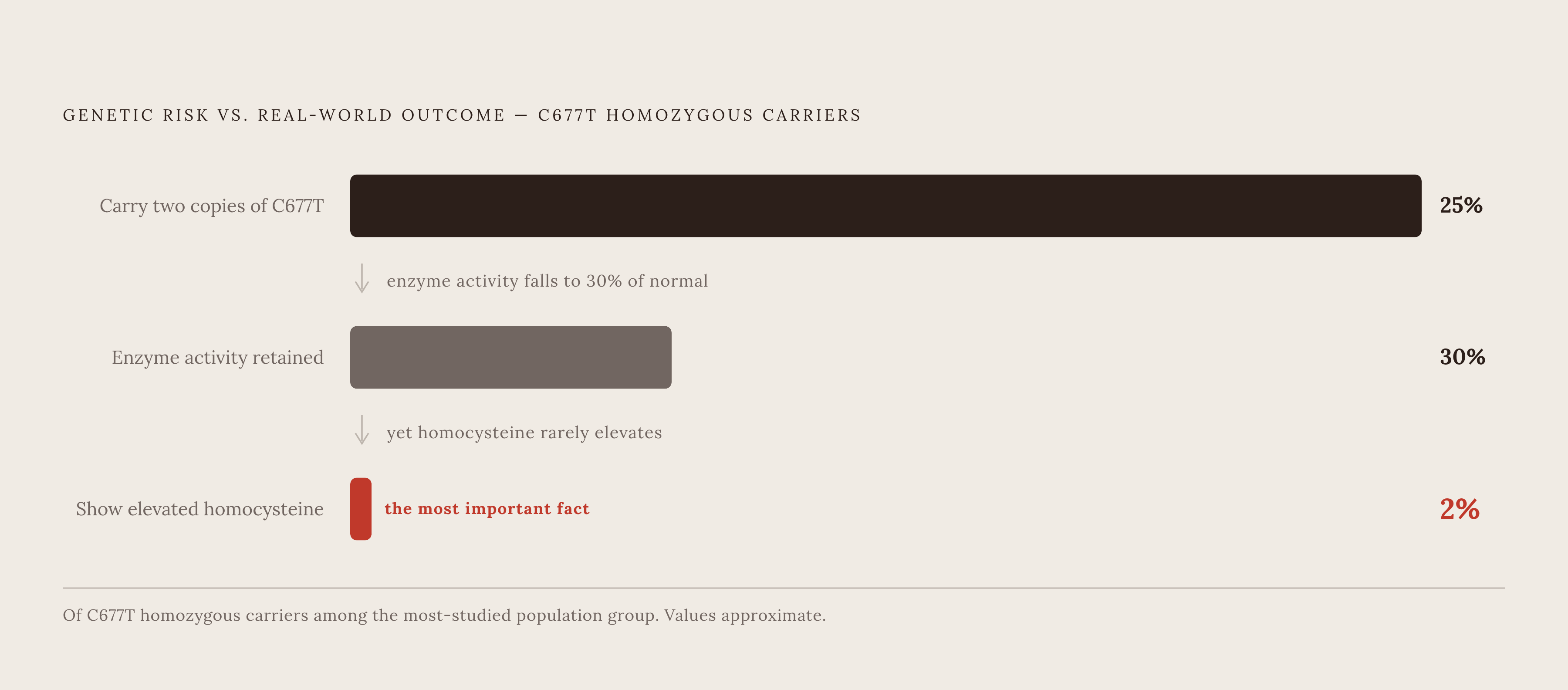
C677T is the common one: cytosine swapped for thymine at position 677. One copy cuts enzyme activity to roughly 65% of normal. Two copies drop it further, to around 30%. On paper, that looks alarming. Roughly 25% of individuals in the population group most studied carry two copies, versus 10 to 15% of Caucasians. Only about 2% of people with two copies of C677T actually show higher-than-normal homocysteine. The enzyme activity drops. The downstream marker, in most people, simply doesn't follow. That gap between genotype and outcome is the single most important fact in this entire topic.
A1298C shows up in roughly 7 to 12% of North American, European, and Australian populations, and the evidence that it meaningfully impairs folate processing on its own is thin. One or two copies doesn't appear to raise homocysteine by itself.
The variant tells you about enzyme capacity. It says nothing about outcome. Treating those as the same thing is where most of the anxiety around MTHFR testing comes from.
The folate testing ecosystem: what serum folate, RBC folate, and homocysteine each measure
Three tests get lumped together under "folate status." They are not interchangeable, and knowing what each one actually captures changes how you should read your own results.
Serum (or plasma) folate reflects recent diet, basically a snapshot of the last few days, and it should be drawn fasting. Below 8 μg/L (18 nmol/L), homocysteine tends to start climbing, a functional warning sign that appears in bloodwork before deficiency would ever show up on a standard blood count. Lab "normal" ranges tend to sit lower than what's actually optimal, too. A serum folate between 10 and 20 ng/mL is a better target, and plenty of people cleared as "normal" on their printout are sitting well under that.
RBC (red blood cell) folate sounds like the more sophisticated test, since it reflects folate stored in red blood cells over roughly three months rather than a few days. It seems like it should tell you something deeper. Does it? A large retrospective analysis comparing a much bigger set of serum samples against a much smaller set of RBC samples found the two track together closely enough that RBC folate rarely catches anything serum folate hasn't already flagged. The College of American Pathologists has gone as far as saying RBC folate no longer meets the standard of care for folate evaluation, precisely because serum folate is more accurate and more reliable. Skip RBC folate. It's the added cost without the added answer.
Homocysteine sits in a different category. It's folate-independent, a functional marker downstream of folate that tells you whether the remethylation pathway described above is actually running the way it should. When it comes back elevated, methylmalonic acid (MMA) becomes the tiebreaker: B12 deficiency raises both homocysteine and MMA, while folate deficiency raises homocysteine and leaves MMA alone. That distinction matters because the two deficiencies get treated differently.
CBC markers (hemoglobin, MCV, MCH, RDW) come last, and they're the least sensitive of the bunch. MCV only drifts above 100, into megaloblastic territory, once deficiency is well established. By the time a CBC shows something, folate and homocysteine have usually been off for a while already.
Why homocysteine is the marker that changes what you do
MTHFR variants don't independently raise the risk of homocysteine-related conditions unless homocysteine itself is actually elevated. Genotype alone doesn't move the needle. It only starts to matter clinically once it appears in bloodwork as a real functional problem.
That flips the priority. A normal homocysteine makes the MTHFR result close to irrelevant, no matter which variant or how many copies someone carries. An elevated homocysteine makes the genotype merit a second look, one piece of a bigger puzzle instead of a headline on its own.
So what counts as elevated? This is where the standard lab range starts to look outdated. Most labs report 5 to 15 µmol/L as normal, wide enough to wave through a lot of people quietly accumulating risk. A meta-analysis from the Homocysteine Studies Collaboration found risk climbing continuously starting above 6 to 7 µmol/L, nowhere close to that 15 µmol/L cutoff. The same analysis tied a 5 µmol/L reduction in homocysteine to a 59% lower stroke risk and a meaningfully lower risk of coronary heart disease. Those aren't small numbers for a marker most people never think about twice.
Functional medicine circles have started using a target below 10 µmol/L, a meaningfully tighter bar than the lab printout suggests. A result of 12 might get filed as "normal" on paper while still sitting in a range worth addressing.
Homocysteine can rise before serum B12 or serum folate themselves test abnormal. It's an early signal, not a late confirmatory one. The upstream nutrient markers often haven't budged yet by the time homocysteine has already told the story.
What drives elevated homocysteine, and how often MTHFR is the reason
Roughly two-thirds of hyperhomocysteinemia traces back to nutrient status: low B12, low folate, or low B6. That single fact should reorder the whole priority list. Diet and supplementation are the first move for most people.
Low B12 is often the primary driver, and here's the odd part: homocysteine tends to rise before serum B12 itself drops low enough to flag. Homocysteine ends up an earlier signal of B12 deficiency than the B12 test itself.
A few other factors push homocysteine up beyond the three vitamins. Declining kidney function is one of the bigger ones, since the kidneys clear homocysteine and a falling GFR lets it build. Smoking, certain medications, and age all nudge it upward too.
MTHFR genotype sits inside that list as one contributing factor. It becomes a real risk factor specifically when serum folate is already low. Once folate status is adequate, the genetic variant tends to fade into the background and stop mattering much.
Homocysteine doesn't only track methylation status, either. It correlates with inflammatory markers like hs-CRP and interleukin-6, so an elevated result is also a marker of systemic inflammation. It's also a hint at systemic oxidative stress, part of why cardiovascular researchers have paid it so much attention over the decades.
What medical guidelines say about MTHFR testing, and what they recommend instead
Given all that, what do the major medical bodies say about testing for MTHFR itself? Both the American College of Medical Genetics (ACMG) and the American College of Obstetricians and Gynecologists (ACOG) recommend against routine MTHFR genetic testing, a fairly firm position from two organizations that don't often issue blanket "don't bother" statements.
Testing gets explicitly discouraged in a specific set of contexts: recurrent pregnancy loss workups and thrombophilia screening, where recent meta-analyses have undercut the supposed link between MTHFR variants and venous thromboembolism risk.
One exception holds up. Certain clinical situations exist where MTHFR status may genuinely inform a treatment decision, and a clinician managing such a case would benefit from knowing it. Outside that lane, the guidance converges on the same alternative every time: skip the genetic test, measure total plasma homocysteine instead. That's the functional readout the guidelines actually want in hand.
How to read your own folate and homocysteine results and respond when something is off
Start simple. Homocysteine plus fasting serum folate makes a practical first panel for anyone curious about methylation status, whether or not an MTHFR result is already sitting in an inbox somewhere. If either comes back off, add serum B12 next. If B12 is in a borderline zone (roughly 200 to 300 pmol/L), holo-transcobalamin and MMA help sort out whether B12 or folate is the actual driver. None of this needs a specialist referral. This kind of check catches a shift before symptoms appear.
Reading the numbers takes a little more nuance than the lab printout offers. Serum folate under 8 μg/L (18 nmol/L) signals depletion, tied to rising homocysteine even before outright deficiency sets in. Serum folate between 10 and 20 ng/mL is the better optimal target than whatever the lab happens to call normal. Homocysteine gets labeled normal anywhere from 5 to 15 µmol/L, but risk climbs continuously above 6 to 7 µmol/L, so a 12 might clear the lab's bar while still deserving a second look.
None of this calls for panicking over a genetic report. A variant is a possibility, not a verdict, and the actual verdict comes from blood, not saliva. Someone with two copies of C677T and a homocysteine of 7 µmol/L sits, functionally, in a different category than someone with that same genotype and a homocysteine of 14. Same genotype, different biology. Test the pathway, not the blueprint.