Cortisol Lab Results and HPA Axis Dysfunction
Testing timing and pattern matter more than a single cortisol number.
A cortisol lab result is a snapshot of a changing system whose handling of stress requires broader assessment. To understand HPA axis dysfunction, it helps to look at when cortisol rises, how it falls, and whether its feedback controls work. The most useful test therefore captures the pattern needed to answer the question.
How the HPA axis produces cortisol and why it does not run at a constant level
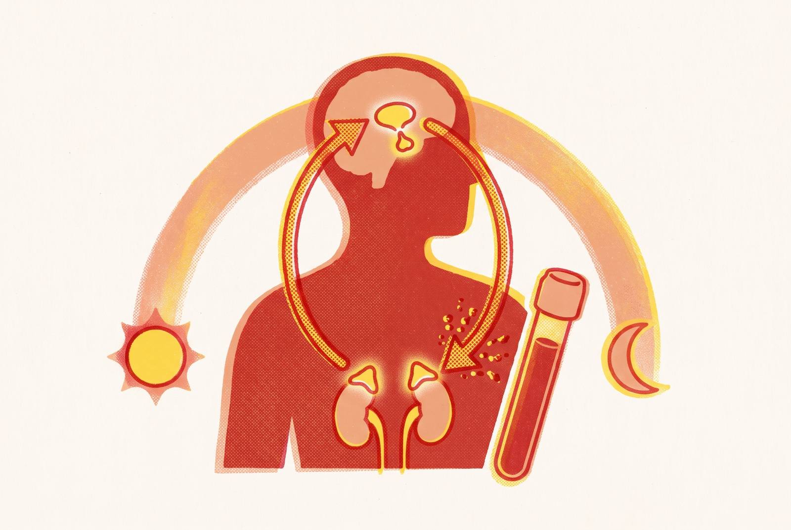
The hypothalamic-pituitary-adrenal axis, or HPA axis, regulates cortisol through a negative-feedback loop. The hypothalamus releases CRH, which signals the pituitary to release ACTH, which in turn signals the adrenal glands to produce cortisol. Each step is a control point. As cortisol rises, it suppresses its upstream drivers so that output can return toward baseline.
This circuit also follows a strong daily rhythm. Cortisol is normally high in the early morning and declines through the day, so collection time is part of the measurement itself. Its metabolic, immune, and cardiovascular work is not harmful in itself. The problem begins when the timing, size, or feedback response of that signal breaks down.
What breaks down when the feedback loop fails
The axis can shift toward prolonged over-activation, suppression, or an oscillating pattern, depending on the stressor, its duration, and individual physiology. In over-activation, cortisol may stay high as the normal drop from morning to evening becomes flatter. Feedback inhibition may also weaken, leaving the upstream signal less responsive to the cortisol already present.
Tomas et al. (2013) described a contrasting pattern in ME/CFS, including low basal cortisol, stronger negative feedback, reduced variation across the day, and a weaker response to challenge. That is a different failure of regulation, not the absence of one. It also explains why interpreting every stress-related symptom as evidence of “high cortisol” can point in the wrong direction.
The same person may show different patterns over time. An initial period of elevated cortisol can be followed by a blunted, low-cortisol state as regulation shifts. This does not mean every person follows that sequence. Changes in methylation of stress-related genes may help some dysregulation last, but researchers are still studying how this works.
Why dysregulated cortisol does not stay confined to stress biology
Cortisol dysregulation can amplify risk well beyond the stress response. Sustained excess can drive insulin resistance, impair glucose metabolism, raise blood pressure, and promote dyslipidemia. Those changes can help cause cardiometabolic disease. Their effects can accumulate before symptoms make the problem obvious.
Impaired feedback and resistance at glucocorticoid receptors can coexist with a pro-inflammatory state, despite cortisol’s role in immune regulation. Cytokine imbalance can then shift immune activity toward autoimmunity. Persistent HPA dysfunction is associated with conditions including systemic lupus erythematosus, rheumatoid arthritis, and multiple sclerosis, although an association does not establish cortisol as their sole cause.
The brain is another site of downstream effects. Chronic glucocorticoid elevation is linked to hippocampal atrophy, disrupted synaptic plasticity, and neuroinflammation, all mechanisms implicated in Alzheimer’s disease pathology. Both high and low cortisol can also impair cognition and emotional stability. Dysregulation that lasts is linked to a greater risk of major depressive disorder and bipolar disorder, while high cortisol over time in children can have lasting effects on how they behave.
A disrupted cortisol pattern together with elevated hs-CRP can reveal an inflammatory dimension that neither result shows alone, providing context for a diagnosis that requires further assessment. To understand the cortisol side of that picture, however, the test must capture the part of the signal under investigation.
What each cortisol test actually measures, and what it cannot
A blood cortisol test answers a narrow question. It measures both protein-bound and free cortisol at the moment of the draw. A morning draw under a defined protocol is the useful starting point for that approach, while a random afternoon value is harder to interpret. Pregnancy, liver disease, and oral contraceptives can alter binding globulins, shifting the result without a matching change in biologically available cortisol.
What if the question is whether cortisol falls normally? Salivary testing measures free, biologically active cortisol, and collection at morning, midday, evening, and bedtime maps the diurnal slope. For assessing that rhythm, a four-point salivary panel is more informative than a single morning blood draw.
The Endocrine Society recommends late-night salivary cortisol as one first-line screening option for suspected cortisol excess. Abnormal results are usually needed from more than one night. Salivary cortisone is also emerging as a stable marker that mirrors serum free cortisol, following conversion of cortisol by 11β-HSD2 in the salivary glands, but it is a different measurement from salivary cortisol.
A 24-hour urine free cortisol test integrates cortisol output across a full day, smoothing moment-to-moment changes, and is a recommended screening test for suspected Cushing’s syndrome. A dexamethasone suppression test instead uses an external glucocorticoid to test whether the axis suppresses appropriately. It challenges the feedback loop rather than simply measuring baseline secretion, so a diurnal saliva panel does not replace it.
Preparation and collection conditions can change all of this interpretation. Steroid medications, including pills, injections, inhalers, and topical creams, are a common source of unexpected cortisol results. A hard blood draw or a rush to get there can also raise serum cortisol for a short time. The practical choice is therefore a test matched to the question, with collection time and medication exposure documented, followed by a comparison of the full cost.
What it costs to test cortisol and how to avoid overpaying
A cortisol test’s price does not reliably indicate its clinical value. Much of the spread reflects insurance-billing arrangements. A single AM serum cortisol test, CPT 82533, can cost less through a direct-access portal even when the collection facility, code, and result are the same. For someone with a high-deductible plan, paying directly may cost less than the share they owe when they use insurance.
As of September 2026, a single blood cortisol test usually costs less than mail-in saliva kits. ZRT Laboratory offers a four-point salivary diurnal panel and a six-point Cortisol Awakening Response panel with DHEAS, identified as S-120 and S-122, at different price points. These are not interchangeable purchases, because a daily slope and an awakening response answer different questions.
Direct-to-consumer ordering can simplify access by sparing someone the need to get a separate physician’s requisition because authorization is part of the order, while leaving interpretation of the result unchanged. To avoid overpaying, the useful comparison is the total charge, including any per-draw or per-order processing fee. An assay advertised at a low price may still come with a higher total bill.
How to read a cortisol result: what patterns signal dysfunction versus normal variation
The first distinction is between an expected peak and persistent elevation. Morning cortisol should be higher than evening cortisol, so “high in the morning” describes part of normal physiology. That is different from dismissing an unexpectedly out-of-range morning result. Interpreting the value requires assessing whether it fits its collection context and whether cortisol then follows the expected decline.
One unusual result is not a complete pattern. Poor sleep, a hard workout, or anxiety during the draw can change one measurement without showing that someone has chronic dysfunction. Day-to-day variation is also normal. An unexpectedly high or low AM serum result should lead to another draw and, ideally, a multi-point salivary panel before a clinician decides what it means.
Cortisol that stays high late at night is more cause for concern. Healthy people usually have bedtime salivary cortisol below 145 ng/dL, and high results on at least two separate occasions call for more checks. A flattened diurnal slope can also suggest over-activation when morning and evening values remain too close together because cortisol does not fall as expected. Failure to suppress after dexamethasone shows that feedback inhibition is impaired, adding to what high output alone can show.
A weak or absent morning peak, followed by low values that change little through the day, suggests a suppressed or hypocortisolic pattern with too little cortisol. The direction of the curve therefore changes its meaning. Symptom history and any preceding period of high cortisol help place that pattern in context.
Additional biomarkers can sharpen the interpretation without settling it alone. DHEA-S falls with age and with prolonged HPA activation, so a low result may help characterize the pattern but must be read in that context. Elevated hs-CRP together with disrupted cortisol strengthens concern about an associated systemic inflammatory state.
Lifestyle and supplement interventions with actual evidence for HPA axis support
Once a problematic pattern is established, the aim is better regulation. How well and how long a person sleeps are the strongest lifestyle foundations because they directly affect the cortisol awakening response and diurnal slope. Disrupted sleep can be both a cause and a consequence of dysregulation. That two-way relationship makes sleep a meaningful input to track.
Exercise also depends on dose and recovery. Moderate aerobic exercise can reduce HPA over-reactivity over time, while excessive training without enough recovery can push toward suppression. Gut microbiome health is another relevant input because dysbiosis can activate immune pathways that interact with the HPA axis. The link runs both ways, so it makes sense to look at the broader system, though this does not show that any particular gut intervention works as a cortisol treatment.
The supplement evidence is more uneven than the lifestyle case. Ashwagandha, including Withania somnifera / KSM-66, has the strongest supplement evidence among the options considered here, with findings of reduced cortisol and anxiety at 8 weeks. In particular, the evidence described here does not establish whether or how often anhedonia occurs, so a lower cortisol result should not be treated as sufficient evidence of improved well-being.
Phosphatidylserine has positive but less generalizable evidence. It acts at the pituitary level to dampen the cortisol signal upstream of the adrenal glands, and findings are most relevant to exercise-induced cortisol responses. Much of that research is smaller-scale, limiting broader conclusions. Magnesium in glycinate or malate forms belongs in a different category, as foundational support for regulation and sleep.
These options are modifiable inputs whose treatment value requires evidence beyond a lab curve. A multi-point panel before and after a targeted intervention can show whether the daily pattern changes, while keeping track of symptoms links the numbers to how the person feels. Comparable collection conditions make that comparison more useful.
Sources
- Chronic Stress and Autoimmunity: The Role of HPA Axis and Cortisol Dysregulation
- The cortisol axis and psychiatric disorders: an updated review
- Hypothalamic-Pituitary-Adrenal (HPA) Axis: Unveiling the Potential Mechanisms Involved in Stress-Induced Alzheimer’s Disease and Depression - PMC
- Chronic Stress-Associated Depressive Disorders: The Impact of HPA Axis Dysregulation and Neuroinflammation on the Hippocampus—A Mini Review
- HPA axis dysregulation is associated with differential methylation of CpG-sites in related genes
- A Review of Hypothalamic-Pituitary-Adrenal Axis Function in Chronic Fatigue Syndrome
- Cortisol Test: What It Is, Purpose, Types & Results
- Waking Salivary Cortisone vs Serum Cortisol in the Short Synacthen Test in Screening for Adrenocortical Insufficiency: Results of a Service Evaluation