TSH Reference Range Controversy and Thyroid Diagnosis
Labs use flawed reference ranges that misdiagnose millions of thyroid patients.
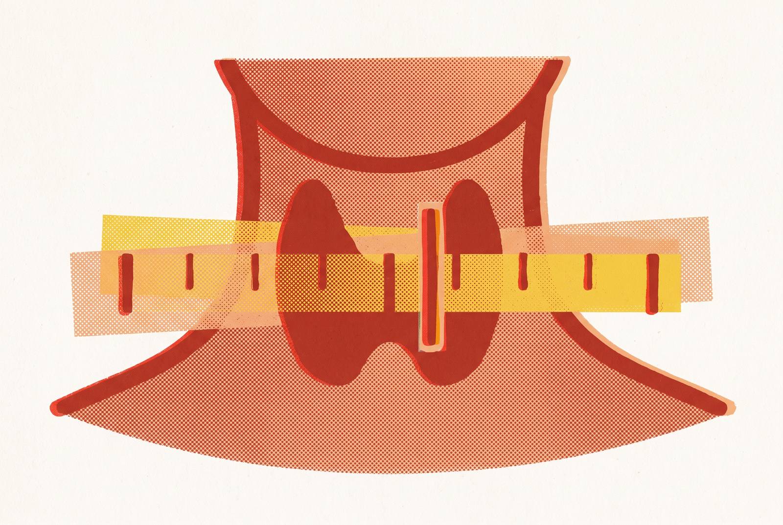
A TSH result marked "normal" on a lab report does not mean a fixed biological line has been crossed or not crossed. It means a number has landed inside a statistical band built from a reference population, and that band is a far shakier foundation than most patients assume.
How a TSH "normal" range is built
Plenty of people have sat across from a doctor, heard "your TSH is normal," and walked out still feeling the fatigue, the brain fog, and the cold hands that sent them in for testing. The confusion makes sense once you see how that "normal" label actually gets made. Labs set the range by taking a reference population and marking off where the 2.5th to 97.5th percentiles fall. Anything outside that band gets called abnormal because statisticians drew a line there, not biology. An ATA-commissioned review of thyroid testing points out that even though TSH is the most ordered hormone test in lab medicine, how to use it well is still an open question, and results can be read more than one way depending on who's reading them. Part of the trouble is that TSH doesn't measure the thyroid directly: it's a signal sent out by the pituitary gland, and it only tells you something true about thyroid function when the whole hypothalamic-pituitary-thyroid loop is working the way it's supposed to. Add to that a mathematical quirk: TSH and free T4 move in a log-linear relationship, so a small, almost unnoticeable drop in free T4 can send TSH climbing out of proportion. That means a tiny shift in where a lab draws its cutoff line can swing a huge number of people from "normal" to "abnormal" overnight, even though nothing changed in their bodies.
The population used to define "normal" TSH includes people with undetected thyroid disease
The upper limit most labs use, somewhere around 4.0 to 4.5 mIU/L, carries a quiet flaw: the "healthy" people used to build that range weren't all healthy. Some had undiagnosed hypothyroidism sitting undetected in their blood work at the time researchers sampled them, and their elevated numbers helped drag the upper boundary higher than it should sit. Some researchers argue that a truly disease-free population would top out closer to 2.5 mIU/L. That number isn't arbitrary. TSH readings above 2.5 mIU/L track with a higher rate of antithyroid antibodies and a greater chance of progressing to full clinical hypothyroidism down the road, which suggests the top end of today's "normal" range already contains early disease hiding in plain sight. The National Academy of Clinical Biochemistry looked at this and stated that the current limits may not reflect a true normal range at all, proposing instead a tighter band of 0.4 to 2.5 mIU/L. The NACB is a credentialed body making a methodological argument, and the stakes of that argument are large: shifting to their narrower range would reclassify somewhere around 22 to 28 million additional Americans. That's how much rides on a decimal point in a reference range. And contamination at the source is only the first problem. A second problem makes the picture worse still, and it comes from something every person experiences: aging.
TSH rises naturally with age, making a fixed upper limit systematically wrong for older adults
TSH climbs as people get older, as a normal part of aging, not as a sign that something has gone wrong. A single fixed ceiling applied to a 30-year-old and an 85-year-old alike is bound to mislabel a large share of older adults as diseased when they're simply aging. NHANES III data put the upper reference limit for TSH at roughly 7.49 mIU/L in adults 80 and older, well above the conventional 4.5 mIU/L cutoff most labs still use for everyone. The clearest demonstration of this comes from a 2025 American Thyroid Association summary of a large Dutch study, built on millions of TSH measurements pulled from numerous institutions. Using age-specific ranges, the researchers found the upper normal limit for TSH in 50-year-old women sat at 4.0 mIU/L, but by age 90 that ceiling had risen by half, to 6.0 mIU/L, while free T4 barely budged across that same stretch of decades. That stability in free T4 alongside a rising TSH points to normal aging physiology driving the TSH shift. Using standard lab ranges as they stand today, a meaningful share of people in their 60s and 70s, and an even larger share past 80, get flagged as abnormal purely because the range wasn't built with their age group in mind. A second, independent population backs this up: one large study of older adults found that mild TSH elevations up to about 5 to 6 mIU/L likely reflect ordinary aging, while levels of 7.0 mIU/L or higher start to carry a real association with worse health outcomes. The French Society of Endocrinology took this problem seriously enough to propose rough arithmetic for adults over 60: take the patient's age in decades, divide by 10, and use that as the upper TSH limit. The formula is simple, almost crude, but it shows a credentialed medical body treating the age problem as something concrete enough to need a fix, not just a footnote.
Sex and race shift TSH baselines further, exposing the "one-size-fits-all" range as a structural problem, not an edge case
Age isn't the only variable the standard range ignores. Sex and race also shape where a person's TSH naturally sits, so the exact same number can mean something different depending on who produced it. When the American Thyroid Association's 2025 analysis applied reference intervals adjusted for age, sex, and race, the reclassification numbers were striking: roughly half of people initially flagged with subclinical hypothyroidism turned out to be normal once personalized ranges were applied, and close to a third of those flagged for subclinical hyperthyroidism were reclassified too. That shift hit older patients, women, and White individuals hardest. The current system applies one range to everyone regardless of these differences, and the same research ties that oversimplification directly to overdiagnosis and underdiagnosis, especially in subclinical cases, where TSH sits outside the range but T4 and T3 look perfectly normal. A result that looks borderline on paper is often, on closer inspection, simply a mismatch between the person being tested and the population the range was built from.
Overdiagnosis from a miscalibrated range leads directly to overtreatment, which carries its own documented harms
Getting the range wrong doesn't just produce a confusing lab report. It pushes real people onto levothyroxine who never needed it, and that drug carries risks you should take seriously. Among women aged 90 to 100 in the Dutch age-specific study, the rate of subclinical hypothyroidism diagnosis would drop from more than one in five down to 8.1% once age-adjusted ranges replace the standard fixed cutoff, a gap wide enough to change treatment decisions for a huge number of people. The harms of overtreating older adults with levothyroxine are well documented: atrial arrhythmias, bone loss, and higher mortality. There's a real possibility that a higher TSH in old age reflects the body adapting, and some researchers have gone as far as proposing that lower T3 in older age might be a marker of longevity. The Korean Thyroid Association ran a 12-year observation study and found that patients with TSH between 4.0 and 6.57 mIU/L showed no adverse health effects over that entire period. Based on that evidence, the KTA set its own upper limit at 6.8 mIU/L, well above the 4.5 mIU/L most labs still use, and the organization strongly recommends against treating mild subclinical hypothyroidism unless TSH climbs above 10 mIU/L. On top of that, the condition often resolves on its own: in roughly three out of five patients with mild subclinical hypothyroidism, TSH returns to normal within five years with no treatment.
The cardiovascular argument for treating borderline TSH is real but age-stratified, and often used without that qualification
The strongest case for treating a mildly elevated TSH rests on cardiovascular risk, and that case holds up, but mainly in younger adults. It weakens substantially when applied to older populations, and it may even flip. Subclinical hypothyroidism has been linked to higher LDL synthesis, more oxidative stress, and impaired function in blood vessel linings, all mechanisms that could plausibly speed up atherosclerosis and strain the heart over time. A 2025 cardiometabolic study in Metabol Open found that people with subclinical hypothyroidism had worse lipid, blood sugar, and liver markers along with higher 10-year cardiovascular risk scores, but treating them with levothyroxine produced only a modest benefit, driven mostly by drops in total and LDL cholesterol. Large studies, including NHANES data and meta-analyses pooling individual patient records, show this cardiovascular link holds up in younger adults, but it fades as people age and TSH naturally climbs higher as part of normal aging. This debate hasn't settled partly for methodological reasons: most of these studies lean on TSH alone as the diagnostic target, and a single number can't tell a thyroid adapting normally from one that's actually failing. The cardiovascular argument deserves to be taken seriously. It just doesn't scale cleanly to every age group the way a single universal range implies it should, and that gap is exactly where the reader's own lived symptoms come back into the picture.
Symptoms at "normal" TSH are not imaginary, they reflect what the single-number model cannot capture
If you land inside the reference range, that doesn't guarantee your thyroid hormone levels are actually adequate for your body. That's why plenty of people with a "normal" TSH still deal with fatigue, weight gain, cold intolerance, thinning hair, and trouble thinking clearly. Multiple studies tie TSH at the high end of "normal" to a modest but measurable uptick in body weight, which gives a real biological mechanism behind a symptom pattern that sometimes gets waved off in a ten-minute appointment. Remember that TSH is a signal sent from the pituitary gland as part of the hypothalamic-pituitary-thyroid axis, and it only works as a stand-in for thyroid health when that whole feedback loop functions correctly. It reflects how the pituitary is responding to circulating hormone, not how much hormone is actually reaching and working in tissue throughout the body. Getting a fuller picture means looking at free T4, free T3, and thyroid antibodies alongside TSH. Free T4 shows how much hormone is actually circulating in the blood. Free T3 shows how much of that hormone the body has successfully converted into its active form. TPO antibodies and thyroglobulin antibodies flag autoimmune thyroid disease, commonly known as Hashimoto's, which can sit there driving real symptoms even while TSH reads perfectly normal. Autoimmune thyroid disease ranks among the most common causes of subclinical thyroid trouble, and a TSH-only panel misses it completely. A panel that skips antibody testing leaves a diagnostic blind spot that no amount of staring at a single TSH number can fill. None of this means a doctor saying "your TSH looks fine" got something wrong. It means TSH by itself is an incomplete signal, one piece of a larger picture that needs more markers to come into focus.
When guidelines disagree on screening and treatment thresholds
Medical organizations themselves haven't settled on who should get screened for thyroid problems, at what age, or what TSH level should trigger treatment, and that open disagreement matters for how patients should approach their own testing. It's a reason to gather complete data and bring real context to any conversation with a doctor. The American Thyroid Association recommends screening starting at age 35. The U.S. Preventive Services Task Force has not recommended routine screening in people without symptoms, citing a lack of solid evidence tying it to better outcomes for cardiovascular disease, illness, or death. The American College of Physicians lands somewhere in between, and it suggests screening for women over 50. Even within a single organization, the ground is shifting: the ATA's 2025 thyroid cancer guidelines moved away from strict numeric TSH targets altogether, replacing them with broader categories like "within normal reference range" for patients free of disease and "below normal reference range" for those with residual disease. Pregnancy offers a useful parallel. Research on TSH reference intervals in pregnant women has settled on trimester-specific ranges as a matter of broad consensus, because one universal cutoff across all three trimesters produces real diagnostic error. If the field accepts that a pregnant woman's TSH needs a different yardstick depending on which trimester she's in, the same logic, that context changes what a number means, applies just as reasonably to age, sex, and race outside of pregnancy. None of this disagreement stems from carelessness. It reflects genuine scientific uncertainty that hasn't been fully resolved. But a patient who waits passively for a referral before ever getting tested may spend years missing context that a fuller panel could have surfaced much sooner.
Accessing a complete thyroid workup and what each marker adds
Given everything a single TSH number fails to capture, a complete thyroid evaluation pulls in five markers together: TSH, free T4, free T3, TPO antibodies, and thyroglobulin antibodies, each one closing a different gap that TSH alone leaves open. TSH still serves as the most sensitive first signal to check, but as this entire discussion shows, its meaning depends heavily on the age, sex, and population context of the person being tested. Free T4 fills in the picture by showing how much thyroid hormone is actually circulating, independent of what the pituitary is signaling, and it tends to stay fairly stable with age. That stability means a rising TSH paired with a steady free T4 is the textbook pattern seen in subclinical cases. Free T3 matters because it measures the active form of the hormone your cells actually use, and when free T3 comes back low while free T4 looks normal, that points to a conversion problem rather than a production problem, something a TSH-only test would never catch. TPO antibodies serve as the main marker for Hashimoto's thyroiditis, and a positive result explains an elevated TSH as autoimmune disease at work. Thyroglobulin antibodies add a second autoimmune marker to round out the picture, and they carry particular value when you monitor patients after thyroid cancer treatment. Getting all five markers checked doesn't require waiting on a doctor's referral or fighting with an insurance company for prior authorization. Direct-pay labs run accredited testing with prices published up front, and a full five-marker panel, covering TSH, free T4, free T3, TPO antibodies, and thyroglobulin antibodies, typically costs a fraction of what the same panel runs through a hospital system. For anyone who has stared at a "normal" result that didn't match how they felt, that full panel is where the real answer usually starts to take shape.
Sources
- What’s New in the 2025 American Thyroid Association’s Guidelines on Thyroid Cancer? - PMC
- Thyroid hormone levels change with age
- thyroid-stimulating-hormone-and- ...
- Indirect reference intervals for TSH in a sample of lebanese pregnant women
- Should there be personalized reference intervals for ...
- Age-specific serum thyrotropin reference range for the diagnosis of subclinical hypothyroidism and its association with lipid profiles in the elderly population
- A Clinical Debate: Subclinical Hypothyroidism - PMC - NIH
- Management of Subclinical Hypothyroidism: A Focus on Proven Health Effects in the 2023 Korean Thyroid Association Guidelines